
Therapeutic ultrasound refers generally to any type of procedure that uses ultrasound for therapeutic benefit.
This includes:
- HIFU
- Lithotripsy
- Targeted ultrasound drug delivery
- Trans-dermal ultrasound drug delivery
- Ultrasound hemostasis, and
- Ultrasound assisted thrombolysis.
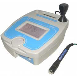
Physical Therapy Ultrasound
Ultrasound Sound is mechanical vibration. The human ear responds to these vibrations in the range 20 Hz to 20 kHz. Sound above 20 kHz is called ultrasound. Therapeutic ultrasound is sound in the range 500 kHz to 5 MHz. Sound waves are produced by some disturbance in a material medium causing the particles or molecules of the medium to vibrate. For this reason sound will not pass through a vacuum.
If the vibration is continuous and regular a constant tone or frequency is produced. The vibration or sound wave propagates through the medium as particles in the medium pass on their vibration to neighbouring particles and series of compressions and rarefactions are produced in the direction of travel of the wave. Therefore, sound waves are longitudinal waves. Sound will travel faster through media where the molecules are closer together and so the velocity is higher in solids than in liquids, and higher in liquids than in gasses.
For example, the velocity of sound in stainless steel is approximately 5800 m/s, in water 1500 m/s and in air only 330 m/s.
As the sound wave passes through the medium, causing molecules to vibrate, some of the energy in the wave is converted from kinetic energy to heat. For a collimated sonic beam the intensity, power per unit area, therefore, decreases exponentially with the distance travelled.
The attenuation of the beam is also dependent upon the frequency of the sound. In solids the attenuation is proportional to frequency, whereas in liquids the attenuation is proportional to the square of the frequency. The usual method of specifying the degree of attenuation of ultrasound in different media is by the half depth. The half depth is the distance the ultrasound must travel through the medium for its intensity to be reduced to one half of its original value.
Many attempts have been made to measure the attenuation in various types of tissue with varying results. It is perhaps more important to remember which types of tissue have the highest absorption and which the lowest. With the lowest absorption first the order is, fat, muscle, skin, tendon, cartilage and bone. For soft tissue the half depth is around 50 mm at 1 MHz and 15 mm at 3 MHz.
It is also important to remember that where there is a change in medium or tissue type there will be both reflection and refraction of the ultrasound beam. In particular, there is almost 100% reflection at the interface of a solid or liquid to air at therapeutic ultrasound frequencies. Any air bubbles in coupling medium will therefore reduce the effective intensity of the ultrasound. Also bone reflects a high percentage of incident ultrasound. It is important, therefore, when applying ultrasound to keep the transducer orthogonal to the surface of the treatment area, to keep the ultrasound transducer moving and to use a good coupling medium to avoid unwanted reflections and locally high intensities.
Therapeutic ultrasound in physical therapy is alternating compression and rarefaction of sound waves with a frequency of >20,000 cycles/second. Therapeutic ultrasound frequency used is 0.7 to 3.3 MHz. Maximum energy absorption in soft tissue is 2 to 5 cm. Intensity decreases as the waves penetrate deeper. They are absorbed primarily by connective tissue: ligaments, tendons, and fascia (and also by scar tissue).
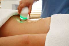
Therapeutic ultrasound may have two types of benefit: Thermal effects and non thermal effects. Thermal effects are due to the absorption of the sound waves. Non thermal effects are from cavitation, microstreaming and acoustic streaming. Cavitational effects result from the vibration of the tissue causing microscopic air bubbles to form, which transmit the vibrations in a way that directly stimulates cell membranes. This physical stimulation appears to enhance the cell-repair effects of the inflammatory response.
Therapeutic ultrasound is sometimes recommended for muscle as well as joint pain.
Contraindications
- Tumours, as ultrasound affects tissue repair and could therefore encourage growth
- Infections, due to the risk of spreading the infection
- Pregnancy, treatment over the pregnant uterus as ultrasound could affect rapidly dividing cells
- Radiotherapy, sites that have received radiotherapy treatment during the last six months
- Thrombosis and impaired circulation.
- Areas of impaired sensation
- Haemorrhage, due to the risk of increased bleeding, including recently controlled bleeding and haematoma.
- Implanted devices such as cardiac pacemakers should be avoided due to the possibility of affecting their operation. Also some plastics used in replacement surgery may be affected by absorption of ultrasound energy. Metal implants may lead to reflections, and as a precaution low doses of ultrasound should be used near these.
- Extreme care should be taken when treating areas near the eye because of the danger of damage to the retina.
- Similarly, extreme care should be taken near the ears and reproductive organs
Ultrasound for drug delivery
Ultrasound has been used in various drug delivery applications to enhance the delivery of pharmaceuticals to target tissues (Acoustic Targeted Drug Delivery). Ultrasound has been shown to facilitate the delivery of drugs across the skin, promote gene therapy to specific tissues, deliver chemotherapeutic drugs into tumours and deliver thrombolytic drugs into blood clots. In addition, ultrasound has also been shown to facilitate the healing of wounds and bone fractures.
Acoustic targeted drug delivery (ATDD) is a method that uses ultrasound energy to enhance the transport of molecules into and/or across specific tissues. Generally this class of ultrasound energy falls under the class of therapeutic ultrasound, and ranges in ultrasonic frequencies of 1-20MHz and Sound intensities of 0-30 watts/cm2. The use of ATDD in conjunction with local drug delivery by injection, topical application and convection enhanced delivery shows promise to significantly enhance the treatment of various diseases in the human body by specifically targeting the drug into the tissue.
ATDD may be applied in the following ways:
- Geometrically, for example with a lens or with a spherically curved transducer.
- Electronically, by adjusting the relative phases of elements in an array of transducers (a "phased array"). By dynamically adjusting the electronic signals to the elements of a phased array, the beam can be steered to different locations, and aberrations due to tissue structures can be corrected.
How ATDD works
Focused ultrasound, similar to high intensity focused ultrasound, but at much lower powers is applied to tissues in conjunction with the local application of a pharmological agent. The ultrasound source (or transducer) is pulsed on and off, and moved in a defined pattern to sonicate the tissue of interest. The pulsing of the ultrasound serves two purposes
- To agitate the tissue matrix by extending and compressing it, and
- To keep the tissue from absorbing to much energy and ablating it (or causing necrosis).

 Left: The manual movement of the transducer allows for control of the sonicated volume of tissue and where the drug may penetrate more readily.
Effective delivery of therapeutic agents into the brain can greatly improve the treatments of neurological and neurodegenerative diseases. Application of focused ultrasound facilitated by microbubbles has shown the potential to deliver drugs across the blood-brain barrier into targeted sites within the brain noninvasively. This review provides a summary of the technological background and principle, highlights of recent significant developments and research progress, as well as a critical commentary on the challenges and future directions in the field. This review also outlines and discusses the tasks that researchers face in order to successfully translate the technology into a clinical reality, including obtaining improved understanding of the mechanisms, demonstration of therapeutic efficacy and safety for specific applications, and development of methodology for rational design to achieve optimized and consistent outcomes.
Therapeutic ultrasound convection enhanced delivery.
Effects of exposure to 1.58 MHz focused ultrasound on transport of drug in soft tissues are enhanced when an external pressure gradient is applied to induce convective flow through the tissue. Drug uptake and transport have been measured in equine brain, avian muscle and agarose brain-mimicking phantoms. Results show that ultrasound enhances drug uptake and transport, and the greatest enhancement occurs when the external pressure gradient is applied. One theory, currently being tested at Cornell, is that exposure of the brain parenchyma to ultrasound enhances penetration of material infused into the brain during convection enhanced delivery therapy.
Therapeutic ultrasound assisted drug delivery to the brain and brain tumours.
Medical treatment for brain tumours may combine radiation therapy with the surgical removal of part of the skull and the excision of the tumour. After resecting the tumour, before replacing the skull section, the surgeon may implant a thin, drug-encapsulated wafer that diffuses chemotherapy agent over time to help ensure that no remaining tumour cells survive.
This approach is too often unsuccessful, and brain cancers like neuroblastomas and neurofibromatosis remain the leading cause of cancer-related death in people under the age of 35. Part of the problem may be that cancerous cells migrate beyond the range of the slowly diffusing drugs.
Therapeutic ultrasound assisted brain drug delivery or Acoustic Targeted Drug Delivery is currently being studied to enhance the success of chemotherapy treatments to brain cancer cells. Scientists are using Therapeutic Ultrasound to increase the distribution of chemical dye agents into brain tissue, to help brain tissue absorb chemotherapy drugs faster. Researchers have found that the use of Therapeutic Ultrasound enhances the chemotherapy delivery and also reduces the time necessary for the drug to work.
It is believed that when focused ultrasound is applied to the brain it agitates the tissues matrices causing enhanced permeability for the drug, and by mechanically pushing it with radiation forces where the acoustical waves are focused. The drugs can then spread further and faster into the tissues than by unassisted diffusion alone. Doctors and scientists hope to use these techniques to increase the effectiveness of chemotherapy as well as reduce the time it takes for the drugs to work in a given patient (e.g., to reach the cancerous brain tissue quickly and before the cells can migrate and regenerate).
Sources:
No comments:
Post a Comment